The summary RAPID calculations show a large region of ischemic at risk parenchyma consisting of the entire brainstem and cerebellum as well as the left occipital lobe. No infarct core is detected by RAPID. There is basilar occlusion as seen on CTA discussed below. If there is any question about whether the TMAX deficit represents simple delayed contrast transit as a result of some upstream stenosis/occlusion that is otherwise chronic and compensated, looking at MTT can help. If MTT is also abnormal (as is the case here) the perfusion deficit is more likely to represent an acute on chronic hemodynamic decompensation.
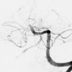
There is occlusion of the basilar artery (red arrow). There is no opacification of the distal right vertebral artery (blue arrow – as it turns out this one is not occluded but just severely narrowed at insertion to the basilar and so the apparent lack of opacification is purely flow related). The left PCA is not clearly opacified compared to the robust right PCA (orange arrows) due to fetal type origin of the right PCA.
No Comments yet!