🎉🎉 New website! We just launched DicomTube, a new powerful platform allowing anyone to anonymize and share full DICOM imaging cases for free! If you find EDNeuroRad helpful, please check us out, sign up and contribute a single case 🙏🏼!
Case 8a Indication: weakness
Loading
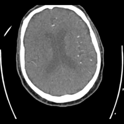
Case 8a: Right MCA stroke
When comparing the right basal ganglia to the left (red dashed area compared to green dashed area), you will notice subtle hypoattenuation with loss of gray-white differentiation. This is typical of an acute infarct within the right MCA territory


No Comments yet!